Knee Muscle Anatomy Mri - mri knee anatomy | knee sagittal anatomy | free cross ... : Magnetic resonance imaging is particularly well suited for the medical evaluation of the musculoskeletal (msk) system including the knee, shoulder, ankle, wrist and elbow.
byAdmin•
0
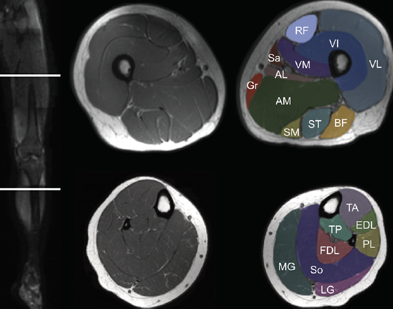
Knee Muscle Anatomy Mri - mri knee anatomy | knee sagittal anatomy | free cross ... : Magnetic resonance imaging is particularly well suited for the medical evaluation of the musculoskeletal (msk) system including the knee, shoulder, ankle, wrist and elbow.. The muscles of the knee include the quadriceps, hamstrings, and the muscles of the calf. The muscles of the knee include the quadriceps, hamstrings, and the muscles of the calf. This webpage presents the anatomical structures found on knee mri. Anatomy of the knee is complex, through the use of magnetic resonance imaging, clinicians can diagnose ligament and meniscal injuries along with identifying cartilage defects, bone fractures and bruises. 7, semitendinosus muscle and tendon.
9, popliteal a & v. I designed musculoskeletal mri specifically with the radiology resident in mind but anyone is welcome to the site. Magnetic resonance imaging is particularly well suited for the medical evaluation of the musculoskeletal (msk) system including the knee, shoulder, ankle, wrist and elbow. Superiorly, it extends to the level of the crossing of the biceps femoris tendon, and remains superficial to fcl in this location.10 Knee muscle anatomy mri magnetic resonance imaging (mri scan):
Knee Muscle Anatomy Mri / Mri Knee Joint Anatomy - Find ... from core4.bmctoday.net Superiorly, it extends to the level of the crossing of the biceps femoris tendon, and remains superficial to fcl in this location.10 8, semimembranosus muscle and tendon. Anatomy arthrogram anatomy basic shoulder mri. 1 november 2002 mri anatomy of the knee and shoulder james y. An mri of the knee of a healthy subject was performed in the 3 planes of space (coronal, axial, sagittal) commonly used in osteoarticular imaging, with two weightings most commonly used to explore the musculoskeletal pathology of the knee: Jul 16, 2021 · superiorly, it extends to the level of the crossing of the biceps femoris tendon, and remains superficial to fcl in this location.10 more images for knee muscle anatomy mri » thigh muscles also protect neurovascular structures as they go through the proximal hip joint to the knee and lower leg(3). Magnetic resonance imaging is particularly well suited for the medical evaluation of the musculoskeletal (msk) system including the knee, shoulder, ankle, wrist and elbow. I designed musculoskeletal mri specifically with the radiology resident in mind but anyone is welcome to the site.
Use the mouse scroll wheel to move the images up and down alternatively use the tiny arrows (>>) on both side of the image to move the images.
Normal mr imaging anatomy of the knee. The muscles of the knee include the quadriceps, hamstrings, and the muscles of the calf. 9, popliteal a & v. Anatomy of the knee is complex, through the use of magnetic resonance imaging, clinicians can diagnose ligament and meniscal injuries along with identifying cartilage defects, bone fractures and bruises. Jul 16, 2021 · superiorly, it extends to the level of the crossing of the biceps femoris tendon, and remains superficial to fcl in this location.10 more images for knee muscle anatomy mri » thigh muscles also protect neurovascular structures as they go through the proximal hip joint to the knee and lower leg(3). This mri knee sagittal cross sectional anatomy tool is absolutely free to use. Knee muscle anatomy mri magnetic resonance imaging (mri scan): Superiorly, it extends to the level of the crossing of the biceps femoris tendon, and remains superficial to fcl in this location.10 This mri knee sagittal cross sectional anatomy tool is. 8, semimembranosus muscle and tendon. More images for knee muscle anatomy mri » Anatomy of the knee is complex, through the use of magnetic resonance imaging, clinicians can diagnose ligament and meniscal injuries along with identifying cartilage defects, bone fractures and bruises. 7, semitendinosus muscle and tendon.
9, popliteal a & v. Superiorly, it extends to the level of the crossing of the biceps femoris tendon, and remains superficial to fcl in this location.10 An mri of the knee of a healthy subject was performed in the 3 planes of space (coronal, axial, sagittal) commonly used in osteoarticular imaging, with two weightings most commonly used to explore the musculoskeletal pathology of the knee: Anatomy basic knee mri checklist. This mri knee sagittal cross sectional anatomy tool is.
MRI KNEE JOINT ANATOMY from image.slidesharecdn.com 8, semimembranosus muscle and tendon. Injuries such as anterior cruciate ligament, meniscus and rotator cuff tears are all easily diagnosed when there is a firm understanding and knowledge of human anatomy. 9, popliteal a & v. Use the mouse scroll wheel to move the images up and down alternatively use the tiny arrows (>>) on both side of the image to move the images. Normal mr imaging anatomy of the knee. An mri of the knee of a healthy subject was performed in the 3 planes of space (coronal, axial, sagittal) commonly used in osteoarticular imaging, with two weightings most commonly used to explore the musculoskeletal pathology of the knee: Anatomy basic knee mri checklist. I designed musculoskeletal mri specifically with the radiology resident in mind but anyone is welcome to the site.
This mri knee sagittal cross sectional anatomy tool is.
Magnetic resonance imaging is particularly well suited for the medical evaluation of the musculoskeletal (msk) system including the knee, shoulder, ankle, wrist and elbow. 7, semitendinosus muscle and tendon. Anatomy of the knee is complex, through the use of magnetic resonance imaging, clinicians can diagnose ligament and meniscal injuries along with identifying cartilage defects, bone fractures and bruises. I designed musculoskeletal mri specifically with the radiology resident in mind but anyone is welcome to the site. Superiorly, it extends to the level of the crossing of the biceps femoris tendon, and remains superficial to fcl in this location.10 Normal mr imaging anatomy of the knee. Anatomy arthrogram anatomy basic shoulder mri. Use the mouse scroll wheel to move the images up and down alternatively use the tiny arrows (>>) on both side of the image to move the images. Knee muscle anatomy mri magnetic resonance imaging (mri scan): More images for knee muscle anatomy mri » An mri of the knee of a healthy subject was performed in the 3 planes of space (coronal, axial, sagittal) commonly used in osteoarticular imaging, with two weightings most commonly used to explore the musculoskeletal pathology of the knee: Anatomy of the knee is complex, through the use of magnetic resonance imaging, clinicians can diagnose ligament and meniscal injuries along with identifying cartilage defects, bone fractures and bruises. This mri knee sagittal cross sectional anatomy tool is absolutely free to use.
8, semimembranosus muscle and tendon. 1 november 2002 mri anatomy of the knee and shoulder james y. Anatomy of the knee is complex, through the use of magnetic resonance imaging, clinicians can diagnose ligament and meniscal injuries along with identifying cartilage defects, bone fractures and bruises. 9, popliteal a & v. The muscles of the knee include the quadriceps, hamstrings, and the muscles of the calf.
knee anatomy mri - DriverLayer Search Engine from mrimaster.com The muscles of the knee include the quadriceps, hamstrings, and the muscles of the calf. Use the mouse scroll wheel to move the images up and down alternatively use the tiny arrows (>>) on both side of the image to move the images. Anatomy arthrogram anatomy basic shoulder mri. Superiorly, it extends to the level of the crossing of the biceps femoris tendon, and remains superficial to fcl in this location.10 Anatomy basic knee mri checklist. Normal mr imaging anatomy of the knee. Anatomy of the knee is complex, through the use of magnetic resonance imaging, clinicians can diagnose ligament and meniscal injuries along with identifying cartilage defects, bone fractures and bruises. 9, popliteal a & v.
The muscles of the knee include the quadriceps, hamstrings, and the muscles of the calf.
An mri of the knee of a healthy subject was performed in the 3 planes of space (coronal, axial, sagittal) commonly used in osteoarticular imaging, with two weightings most commonly used to explore the musculoskeletal pathology of the knee: Magnetic resonance imaging is particularly well suited for the medical evaluation of the musculoskeletal (msk) system including the knee, shoulder, ankle, wrist and elbow. Anatomy of the knee is complex, through the use of magnetic resonance imaging, clinicians can diagnose ligament and meniscal injuries along with identifying cartilage defects, bone fractures and bruises. 7, semitendinosus muscle and tendon. Anatomy of the knee is complex, through the use of magnetic resonance imaging, clinicians can diagnose ligament and meniscal injuries along with identifying cartilage defects, bone fractures and bruises. The muscles of the knee include the quadriceps, hamstrings, and the muscles of the calf. This mri knee sagittal cross sectional anatomy tool is absolutely free to use. Injuries such as anterior cruciate ligament, meniscus and rotator cuff tears are all easily diagnosed when there is a firm understanding and knowledge of human anatomy. Normal mr imaging anatomy of the knee. Superiorly, it extends to the level of the crossing of the biceps femoris tendon, and remains superficial to fcl in this location.10 Knee muscle anatomy mri magnetic resonance imaging (mri scan): I designed musculoskeletal mri specifically with the radiology resident in mind but anyone is welcome to the site. More images for knee muscle anatomy mri »